Limitation in movement of the fourth Finger.
A source of constant trouble to the piano teacher is the limitation in movement of the fourth finger. This results, primarily, from the presence of ligamentous bands connecting the tendon of this finger with that of the third and that of the fifth.
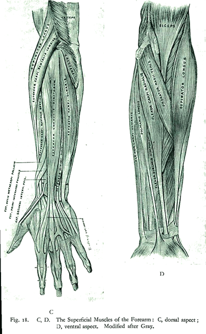
These bands are shown in Fig. 18 C. (See below) Their position indicates that they limit extension of the finger, not flexion. That is, they affect finger lift, not the down stroke. No amount of practice can overcome entirely this physiological limitation. What practice does is to extend the band very slightly and to increase the force of the fourth finger stroke, thus making less lift necessary for the production of a tone of given intensity. The fourth finger never reaches the independence of the others.
The following figures give the distances through which each finger, after prolonged drill in extending this range, could be lifted in a particular case. The hand was held in a normal position for playing, and as each finger was lifted, the remaining fingers remained in contact with the key surface. The distances given are the vertical distances between the finger-tip and the surface of the piano-key.
Right Hand: Second finger, 3'25" ; third finger, 2'80" ; fourth finger, 1'60" ; fifth finger, 2'50".
Left Hand: Second finger, 3'25'; third finger, 2'80' ; fourth finger, 1'50'; fifth finger, 2'50'.
Left Hand: Second finger, 3'25'; third finger, 2'80' ; fourth finger, 1'50'; fifth finger, 2'50'.


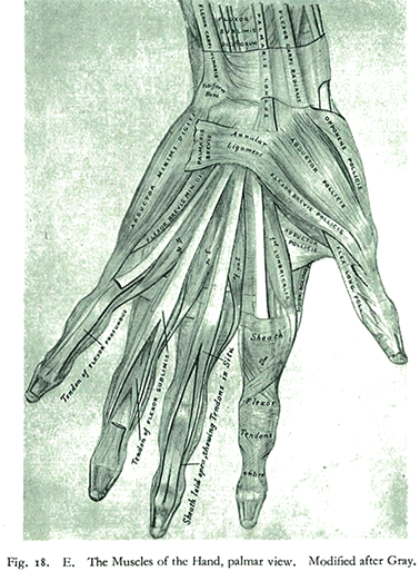
Training thus changes the absolute amount of finger lift but not its relation to that of the other fingers.Finally the presence of two small muscles explains the greater lift for the second and the fifth fingers. Each of these digits has an additional extensor, helping to increase its range of lift. The subdivisions of the hand, illustrated in Fig. 165, showing the isolation of fifth and second fingers, are thus seen to have a muscular cause. (The two muscles in question are the extensor indicis [1] and the extensor minimi digiti.) (See Fig. 18, A, B, C, D.)
Recap: Muscules of the hand
The muscles of the hand are subdivided into three groups:
- those of the thumb, which occupy the radial side and produce the thenar eminence;
- those of the little finger, which occupy the ulnar side and give rise to the hypothenar eminence;
- those in the middle of the palm and between the metacarpal bones.
[1]extensor indicis: The extensor indicis [proprius] is a narrow, elongated skeletal muscle in the deep layer of the dorsal forearm, placed medial to, and parallel with, the extensor pollicis longus. Its tendon goes to the index finger, which it extends.