Muscles of the Thumb
- The movements of the thumb are performed by eight muscles.
- Four of the muscles are situated in the forearm and four in thenar eminence. [1]
Of the four muscles in the forearm three are extensors, one for each thumb-joint. The action, however, is not entirely isolated. One of these extensors (longus pollicis) extends the thumb tip, but if the movement be continued it acts also upon the other thumb-joint. This extensor is attached to the bone differently from the similar tendon in the other fingers. Accordingly, the impossibility of extending the nail-joint while flexing the other joints does not hold for the thumb. As in the case of all the muscles thus far considered, the muscles of the thumb show clearly the integrative action of the muscular system.
- The short extensor of the thumb ordinarily affects the first phalanx.
- As the movement increases in force the whole thumb is pulled back and the wrist muscles are innervated to prevent the movement from spreading to the wrist.
- The short flexor of the thumb flexes the nail joint and helps to move the entire thumb toward the little finger.
- The chief abductor extends the nail joint, moves the middle phalanx sideways, and abducts the entire thumb.
- A combination of these functions (although opposite in kind) are also found in the thumb-abductor.
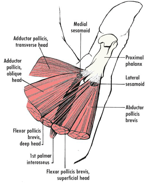
- Flexor pollicis brevis, superficial head: The Flexor pollicis brevis, superficial head is a muscle located in the thenar eminence (the fleshy mound at the base of your thumb). Its primary function is to flex the thumb at the metacarpophalangeal joint (the joint where the thumb connects to the hand), allowing you to perform actions like gripping and pinching.
- first palmar interosseous: The first palmar interosseous is a short, unipennate skeletal muscle in the hand's interosseous compartment. It's also known as the pollical palmar interosseous and is located on the medial side of the thumb
- Flexor pollicis brevis, deep head: The Flexor pollicis brevis, deep head is a small muscle located deep within the base of the thumb. Its primary function is to flex the thumb at the metacarpophalangeal joint (where the thumb meets the palm), enabling powerful gripping and pinching actions.
- adductor pollicis, oblique head: The adductor pollicis, oblique head muscle is a key muscle in the thenar eminence (the fleshy area at the base of the thumb). Its primary functions are to draw the thumb towards the palm (adduction) and assist in rotating the thumb inwards, enabling powerful pinching and gripping movements.
- adductor pollicis transverse head: The adductor pollicis transverse head is one of the two parts of the adductor pollicis muscle in the thumb. It arises from the palmar base of the third metacarpal bone and inserts into the ulnar side of the base of the proximal phalanx of the thumb. Along with the oblique head, the transverse head helps pull the thumb inwards towards the palm . This action is crucial for gripping and manipulating objects.
- medial sesamoid bone: The medial sesamoid bone of the thumb is embedded within the adductor pollicis muscle. Together with the lateral sesamoid bone (in the flexor pollicis brevis), they act to increase the mechanical advantage of the thumb flexor tendons, enhancing power and precision in grip and pinch movements.
- Proximal Phalanx: The proximal phalanx muscles of the thumb are responsible for flexing the thumb at the metacarpophalangeal (MCP) joint, allowing you to bend your thumb towards your palm. These muscles are essential for gripping objects and performing fine motor tasks.
- lateral sesamoid bones: The lateral sesamoid bones of the thumb are embedded within the tendons of the short thumb muscles (abductor pollicis brevis, flexor pollicis brevis). These small bones act as pulleys, improving the leverage of the thumb muscles and protecting the tendons from wear and tear.
- adductor pollicis brevis: The adductor pollicis brevis muscles are a group of small muscles located at the base of the thumb, within the thenar eminence. Their primary function is to adduct the thumb, which means drawing the thumb inward towards the palm for actions like gripping and pinching.
Anatomy, Purpose and Function of the Thumb Muscles
The thumb muscles are a specialized group of muscles responsible for the thumb's incredible range of motion, strength, and dexterity. These muscles are essential for grasping, pinching, and manipulating objects, making the thumb a vital part of human hand function. Here's an overview of their anatomy, purpose, and function:
Anatomy of the Thumb Muscles
The muscles associated with the thumb can be categorized into two groups based on their location: a) intrinsic muscles (within the hand) and b) extrinsic muscles (originating from the forearm).
Purpose of the Thumb Muscles
The thumb muscles serve to:
Functions of the Thumb Muscles
The combined actions of the thumb muscles allow for:
Clinical Relevance
By orchestrating complex and coordinated movements, the thumb muscles are central to the hand's versatility and utility in daily life.
Anatomy of the Thumb Muscles
The muscles associated with the thumb can be categorized into two groups based on their location: a) intrinsic muscles (within the hand) and b) extrinsic muscles (originating from the forearm).
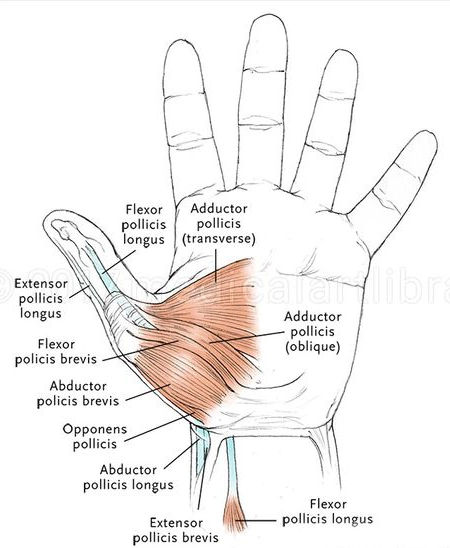
- Intrinsic Thumb Muscles (Thenar Muscles)
Located in the thenar eminence (the fleshy part of the palm near the thumb), these muscles are responsible for fine motor control and precision movements.
-
Abductor Pollicis Brevis:
- Location: Superficial and lateral in the thenar eminence.
- Function: Abducts the thumb (moves it away from the palm), enabling opposition and grasping.
-
Flexor Pollicis Brevis:
- Location: Medial to the abductor pollicis brevis.
- Function: Flexes the thumb at the carpometacarpal and metacarpophalangeal joints.
-
Opponens Pollicis:
- Location: Deep to the abductor pollicis brevis.
- Function: Allows the thumb to oppose the fingers by rotating and flexing the carpometacarpal joint.
-
Adductor Pollicis:
- Location: In the web of the hand between the thumb and index finger.
- Function: Adducts the thumb, pulling it toward the palm for a strong grip.
-
Abductor Pollicis Brevis:
- Extrinsic Thumb Muscles
These muscles originate in the forearm and extend into the hand to control thumb movement.
Here is the HTML unordered list with the child elements nested into another unordered list:
-
Flexor Pollicis Longus:
- Location: Deep in the forearm, tendon runs to the thumb.
- Function: Flexes the thumb, particularly at the interphalangeal joint.
-
Extensor Pollicis Longus:
- Location: Posterior forearm.
- Function: Extends the thumb at the interphalangeal and metacarpophalangeal joints.
-
Extensor Pollicis Brevis:
- Location: Adjacent to the extensor pollicis longus.
- Function: Extends the thumb at the metacarpophalangeal joint.
-
Abductor Pollicis Longus:
- Location: Posterior forearm.
- Function: Abducts the thumb and assists in extension.
-
Flexor Pollicis Longus:
Purpose of the Thumb Muscles
The thumb muscles serve to:
- Provide the thumb with an unparalleled range of motion, including opposition, flexion, extension, abduction, and adduction.
- Facilitate precision grip (e.g., pinching) and power grip (e.g., grasping).
- Support fine motor tasks, such as writing, buttoning, and using tools.
Functions of the Thumb Muscles
The combined actions of the thumb muscles allow for:
-
Opposition:
- The thumb moves across the palm to touch the fingertips, enabling grasping and manipulating objects.
-
Pinch Strength:
- Movements such as holding a pen or picking up small objects rely on the coordination of the intrinsic and extrinsic thumb muscles.
-
Grasping Power:
- Adduction and flexion of the thumb enhance grip strength for larger or heavier objects.
-
Stabilization:
- Muscles like the adductor pollicis stabilize the thumb during activities requiring sustained pressure.
-
Dexterity:
- The thenar muscles enable intricate movements needed for tasks like typing, playing musical instruments, and sewing.
Clinical Relevance
- Injuries or conditions such as carpal tunnel syndrome, De Quervain's tenosynovitis, or arthritis can impair thumb muscle function, leading to reduced grip strength and dexterity.
- Rehabilitation and strengthening exercises often focus on these muscles to restore function after injury.
By orchestrating complex and coordinated movements, the thumb muscles are central to the hand's versatility and utility in daily life.
Origin and Insertion of Muscles
The attachment of the muscular tendon to the rest-end is called the origin of the muscle. The tendonous attachment to the moveable bone is called the insertion. Between the origin and insertion of a muscle at least one articulation must intervene. In the case of several muscles, more than one joint intervenes. The entire problem of muscular action depends upon the relationship between muscle position and joint. The question as to which end of a muscle is its origin and which is its insertion cannot always be definitely answered until the movement is seen. Muscular contraction through the principle of the opposite equality of forces will exert a pull on both bones to which the muscle is attached. The bone offering the less resistance will be the one to move. Origin and insertion may change places. The origin of a muscle will be the end closer to the trunk. [2]
[1]thenar eminence: The thenar eminence ("palm of the hand") and the Latin word "eminentia", meaning projection) refers to the group of muscles on the palm of the human hand at the base of the thumb.
[2]longus pollicis: a muscle that arises dorsolaterally from the middle part of the ulna, extends the second phalanx of the thumb, and abducts the hand.
[3]trunk: n. [ME. tronke < OFr. tronc < L. truncus, a stem, trunk < truncus, maimed, mutilated < IE. *tronkus < base *trenk-, to press together, crowd, whence THRONG]
the main body or stem of a nerve, blood vessel, muscle, as distinguished from its branches.
the main body or stem of a nerve, blood vessel, muscle, as distinguished from its branches.